3D Imaging in Dentistry

KatieM

Katie Moynihan, BS RDH

3D Imaging in Dentistry

Dental x-rays are a routine part of your dental visit. Unfortunately, x-rays can only show the healthcare provider a 2D image of your tooth structure and supporting bone. Our North Stapley office is excited to now offer our patients a 3D imaging device called Cone Beam Computed Tomography, or CBCT. All patients at any of our locations can utilize this great technology.  This device is able to capture a 3D scan of a patient’s maxillofacial skeleton for diagnostic purposes.

Uses for a CBCT Scan:

CBCT scans are used in many different fields of dentistry to improve diagnosis and treatment planning in the following cases:

Endodontics
-Tooth morphology, number of canals and root curvature
-Identification of periapical pathology
-Location of trauma, root fractures

Dental implants
– Location of anatomic structures
– Size and shape of ridge, quantity and quality of bone
-Number, orientation of implants
-Need for bone graft, sinus lift
– Use of implant positioning software

Oral and maxillofacial surgery
– Relationship of third molar roots to mandibular canal
– Localization of impacted teeth, foreign objects
-Evaluation of facial fractures
-Location and characterization of lesions

Orthodontics
– Treatment planning for complex cases
– Impacted teeth
-Root angulation, root resorption

Sleep Apnea
-Identification of obstructive airway

Temporomandibular joint or TMJ
– Osseous structures of TMJ
-Relationship of condyle and fossa

Screen Shot 2015-10-25 at 9.59.31 PM

How Does It Work:

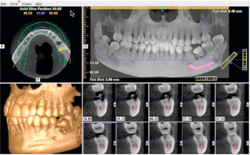
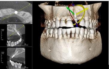
The patient is precisely placed in a comfortable position at the machine. The scan takes about 20 seconds to rotate around the head, obtaining nearly 600 distinct images. The focused x-ray beam reduces scatter radiation, resulting in better image quality. Once complete, the 3D image is immediately available for viewing and diagnosing. The scan produces a wide variety of views and angles that can be manipulated to provide a more comprehensive evaluation. One CBCT scan uses about 1/20th the radiation of a traditional head and neck scan at the hospital.

Screen Shot 2015-10-25 at 10.14.56 PM

There are many benefits to using a 3D imaging CBCT system in dentistry. We are excited to be able to provide top of the line technology to our patients. This new machine will be beneficial in increasing predictability by decreasing failure to provide you with the best quality of care!

Screen Shot 2015-10-25 at 10.15.10 PM

Want to learn more? Visit us at

http://www.shalimarfamilydentistry.com

http://www.northstapleydentalcare.com

http://www.alamedadentalaz.com

http://www.dentistingilbert.com

Sources:


http://www.fda.gov/Radiation-EmittingProducts/RadiationEmittingProductsandProcedures/MedicalImaging/MedicalX-Rays/ucm315011.htm

http://www.conebeam.com/whatis

http://www.radiologyinfo.org/en/info.cfm?pg=dentalconect#benefits-risks


Tooth Anatomy

KO6A3321-Edit

Becky Larson, RDH

Tooth Anatomy

Sometimes when I am talking to my patients I can tell they are a bit confused!  I never want my patients to be confused.  Communication is so important when trying to give important information about oral health.  This blog is just a quick overview of the anatomy of the tooth.  Knowing a few simple terms can eliminate confusion and patient’s can better understand why their dentist and hygienist are giving specific recommendations for home care and dental treatment.

Tooth Anatomy

This diagram breaks the tooth into two parts: the crown and the root.  The root surface of the tooth is the part that is covered by gum tissue in a healthy mouth.   The crown of the tooth is that part we actually see.  Sometimes the root of the tooth is exposed due to gum recession and can be sensitive to cold or vibration.  The gums or gingival cover the jaw bone in which the teeth sit.

Teeth are composed of several layers.  The outer most layer is the enamel, which is generally white in color.  Though it is the hardest substance in the human body, it can still decay and erode if not taken care of.  Just under the enamel is the dentin which is much softer and more yellow in color.  When viewing radiographs, if the dentist sees a cavity that has progressed through the entire enamel layer and into the dentin, a filling is necessary.

Radiograph (xray) showing a cavity that has progressed through the enamel into the dentin:

Decay

Under the dentin layer is the pulp.  There are also nerves and blood vessels in the pulp chamber that extend down the roots of the tooth.  If a cavity has progressed into the pulp chamber, it’s likely that a root canal is necessary.  During the process of a root canal, the nerves and pulp inside the canals are taken out.  The inside of the tooth is cleaned and sealed to prevent further pathology.

Radiograph showing tooth roots after a root canal:

Root Canal

Understanding tooth anatomy can help patients better understand their dental needs!  Always consult your dentist or dental hygienist if you have concerns or questions about your teeth or oral health.

Want to learn more? Visit us at

http://www.shalimarfamilydentistry.com

http://www.northstapleydentalcare.com

http://www.alamedadentalaz.com

http://www.dentistingilbert.com

Sources:

https://www.humana.com/learning-center/health-and-wellbeing/healthy-living/tooth-enamel

http://www.webmd.com/oral-health/guide/dental-root-canals

What is a Dental Porcelain Veneer?

LindsayW

Lindsay Olsen, RDH

What is a Dental Porcelain Veneer?

Dental veneers (sometimes called porcelain veneers or dental porcelain laminates) are wafer-thin, custom-made, shells of tooth-colored materials designed to cover the front surface of teeth to improve your appearance.  These shells are bonded to the front of the teeth changing their color, shape, size, or length.

Screen Shot 2015-10-11 at 7.43.34 AM Screen Shot 2015-10-11 at 7.43.44 AM

Screen Shot 2015-10-11 at 7.43.52 AM Screen Shot 2015-10-11 at 7.44.05 AM

Screen Shot 2015-10-11 at 7.44.14 AM

Screen Shot 2015-10-11 at 7.44.41 AM

In 2011, following the removal of my braces, Dr. Jason Nelson placed two all porcelain veneers on my front upper teeth. Below are pictures of my own teeth.

Screen Shot 2015-10-11 at 7.46.27 AM

Screen Shot 2015-10-11 at 7.46.40 AM

Please contact your dental office for more information regarding dental veneers!

http://www.alamedadentalaz.com/

http://www.northstapleydentalcare.com/

http://www.shalimarfamilydentistry.com/

http://www.dentistingilbert.com/

Images:

https://www.google.com/search?q=what+is+a+dental+veneer&biw=1600&bih=731&source=lnms&tbm=isch&sa=X&sqi=2&ved=0CAYQ_AUoAWoVChMIpu_Mq-y4yAIVF-1jCh2gjgyj#imgrc=-Dp20x_5-7xrrM%3A

https://www.google.com/search?q=what+is+a+dental+veneer&biw=1600&bih=731&source=lnms&tbm=isch&sa=X&ved=0CAcQ_AUoAWoVChMI0sXxp-24yAIVQ8hjCh2sSgh6#tbm=isch&q=what+is+a+dental+porcelain+veneer&imgrc=WoNzbSiOtXK1EM%3A

https://www.google.com/search?q=what+is+a+dental+veneer&biw=1600&bih=731&source=lnms&tbm=isch&sa=X&ved=0CAcQ_AUoAWoVChMI0sXxp-24yAIVQ8hjCh2sSgh6#tbm=isch&q=what+is+a+dental+porcelain+veneer&imgrc=hACaPJFsiKsG-M%3A

A little dental humor to keep you smiling

KO6A8495-Edit - Copy

Lora Cook, RDH

A little dental humor to keep you smiling…

Flossing

Flossing More

AirFloss

You can always air floss!

Loose Canine

For more fun, Visit us at

http://www.shalimarfamilydentistry.com

http://www.northstapleydentalcare.com

http://www.alamedadentalaz.com

http://www.dentistingilbert.com

Sources:

http://www.sugarfixdental.com/

http://www.viralnova.com/funny-dog-faces/

http://www.buzzfeed.com/ailbhemalone/things-youll-only-know-if-your-parents-were-dentists?sub=2681816_1827186

http://themetapicture.com/must-visit-the-dentist/